qEEG brain mapping –
measure first, then treat
Not every patient with us gets a qEEG, and that is deliberate. Everyone starts with a structured assessment: history, existing findings, questionnaires, clinical examination and, where needed, lab work and imaging. qEEG brain mapping is added when the question calls for it, for instance before neurofeedback or when it is unclear which network sits behind the symptoms; then it shows, with 32 channels, what a description of symptoms alone cannot.
qEEG stands for quantitative EEG. We record your brainwaves and analyse them computationally: frequency bands, power and connectivity are measured, displayed as colour topographic maps (“brain maps”) and compared with a normative database. qEEG is a diagnostic and orientation tool, not a therapy, and it does not replace a medical diagnosis; it gives the physician an additional, objective piece of information. Who gets it: people for whom we plan neurofeedback, people whose choice of method depends on the network profile, or people whose symptoms and findings do not add up. Who does not need it: someone with a clear indication whose history, examination and existing findings already carry the treatment. A measurement that changes no decision is one we leave out.
Our device: Neuroelectrics Enobio 32 (32-Kanal-Wireless-EEG)
The mechanism, made clear
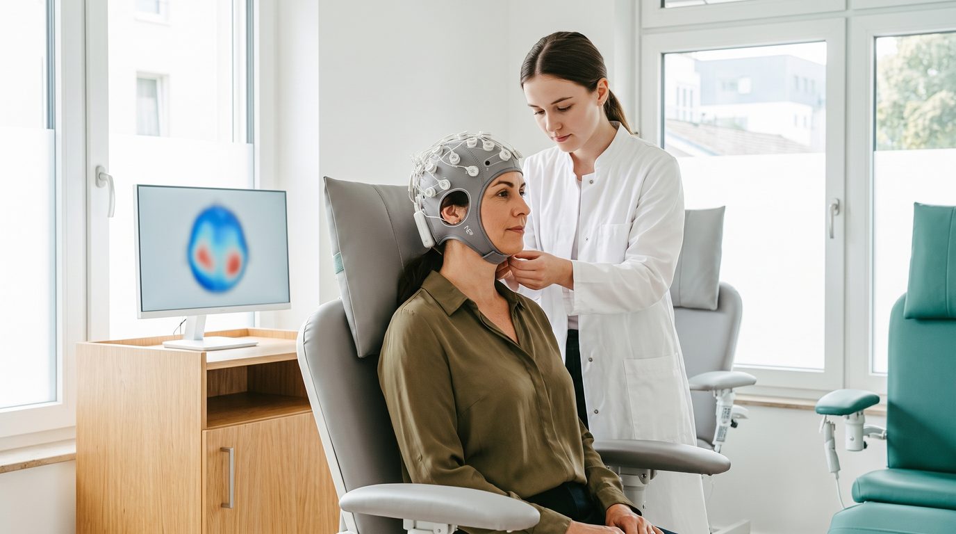
Recording is done with the Neuroelectrics Enobio 32, a 32-channel wireless EEG with a light electrode cap and a small NECBOX recording unit. At rest, with eyes open and closed and, where needed, during tasks, activity is captured at many scalp locations; the software computes frequency and connectivity measures from it and sets them against normative values. A qEEG on its own explains no symptom. A slowed frontal rhythm can be an underactive thyroid, a restless salience network an inflammation coming from the gut or from an infection; that is why the physician reads the maps next to labs, heart rate variability, imaging and history, and the assessment decides beforehand whether the map is needed at all.
qEEG · Alpha 8–12 Hz · relative power
What to expect
The measurement is painless and non-invasive: you wear the cap, sit still, and open and close your eyes on cue; recording takes 20 to 40 minutes. Nothing is delivered into the brain; the qEEG only listens. No radiation, no contrast agent, no side effects; afterwards we go through the findings with you. What the maps show us in practice: two people with the same diagnosis rarely show the same pattern, and the same person shows different ones after a good night and after a bad one. So when we measure, we measure under defined conditions and never interpret a map on its own; and what we learn from the interplay of map, labs and course changes the next concept we build for someone.
Where the question calls for it, qEEG brain mapping is the diagnostic building block of our program: it makes the network profile visible and shows which methods fit it, such as neurofeedback, rTMS or other neuromodulation. It is used, among other things, in ADHD, depression, in cognitive questions and for follow-up, when there is a baseline to compare against.
See all conditions — and what fits you is decided by the assessment, with qEEG where the question calls for it.
What the research shows
Peer-reviewed work on qEEG Brain Mapping — graded by evidence level. No study proves an individual outcome.
The value of quantitative electroencephalography in clinical psychiatry: a report by the Committee on Research of the American Neuropsychiatric Association
Quantitative electroencephalography as a complement to symptom-based psychiatric diagnosis: a narrative review
Conventional and quantitative electroencephalography in psychiatry
The quantitative EEG theta/beta ratio in ADHD and normal controls: sensitivity, specificity, and behavioral correlates
Quantitative EEG in children and adults with ADHD: comparison of absolute and relative power spectra and theta/beta ratio
Quantitative EEG in the differential diagnosis of dementia subtypes
Quantitative electroencephalography in the diagnosis of dementia with Lewy bodies
The neurobiology of the EEG biomarker as a predictor of treatment response in depression
EEG biomarkers to predict response to sertraline and placebo treatment in major depressive disorder
Traumatic brain injury: an EEG point of view
Quantitative EEG signatures of power and functional connectivity alterations in Alzheimer’s disease and frontotemporal dementia
More: Science & Studies.
Professional bodies classify qEEG as an objective adjunct whose single measures, such as the theta/beta ratio, become meaningful only together with the clinical picture and the other findings. With us it is therefore recorded when it changes a decision, and then read by a physician.
Part of Clinicum St. Georg, under the medical direction of Dr. med. univ. Julian Douwes. Outpatient, evidence-graded, tailored to your profile. More on our program.
qEEG Brain Mapping: is it right for you?
The assessment decides, not a brochure. We clarify it in a free consultation, with no obligation and full confidentiality.